
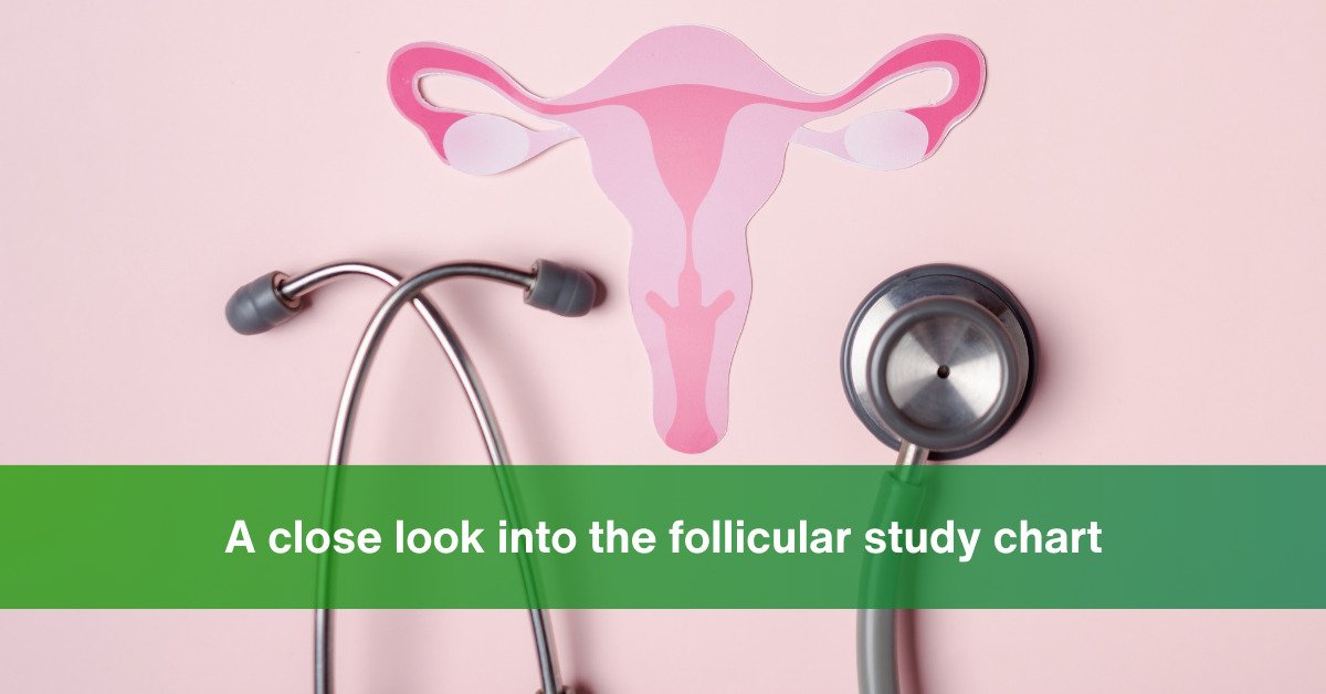
There are many who say, “I have the Follicular report in my hand, but I don’t know what exactly the Follicular study chart tries to convey!” If you are one among them, then this blog is for you.
We will run through the intricacies of the Follicular Study Chart, inch by inch for you, with reference to our detailed report at Jammi Scans.
What is a follicular analysis?
A series of scans that are performed to determine when the mature egg will be released and the uterine walls will thicken is follicular analysis. This analysis is crucial to your pregnancy journey, especially if you are trying for a baby.

The follicular analysis monitors,
- The number of follicles
- The changes in the uterine lining
- The ovulation period so that the intercourse/IUI/IVF procedure can be timed appropriately
- If there is a need to adjust the dosage of any prescribed fertility medications
- Detects follicle growth issues or other ovulation issues
What is a follicular study chart?

A follicular study chart is a mere representation of the follicular analysis done by the fetal medicine specialist. It has certain metrics based on which the study happens to learn the development of follicles in a woman’s ovaries.
Some of the crucial pointers that the follicular study chart tracks are mentioned in detail below:
The date and the day of the cycle:
This column has two dates – one is the first day of your period and the second is the start date of the follicular study.
ET column:
The second column is called the ET otherwise the Endometrium Thickness. Your fetal medicine specialist tracks the Endometrium Thickness at every sitting of your follicular study here.
Note: As the cycle progresses toward ovulation, the endometrium thickens to about 11 mm. An egg is released 14 days into a person’s cycle due to hormones. Endometrial thickness is at its peak during this secretory phase and can reach 16 mm.
Endometrial thickness is significant during pregnancy. The best chances for a healthy, full-term pregnancy, according to medical experts, are associated with an endometrium that is neither too thin nor too thick.
As a result, the embryo can successfully implant and get the nutrition it needs, gradually thickening the endometrium as the pregnancy progresses.
The most common method for measuring endometrium thickness is ultrasound. It is the first method used by healthcare providers, especially when an individual reports abnormal vaginal bleeding.
Doctors use MRI when ultrasound is inappropriate, which is often due to the position of a person’s uterus or other health conditions.
Right Ovary and Left Ovary:
This column tracks the MSF or the Multiple Small Follicle presence and its sizes in detail. As the week progresses, the study tracks the trace of the dominant follicles, the HCG injection given, whether it is collapsing or ruptured in the state.
Note:
The ovaries are roughly the size and shape of an almond and sit just above the fallopian tubes—one on each side of the uterus. Every month during ovulation, either the right or left ovary yields a mature or the dominant follicle for fertilization in a fertile female.
When the dominant follicle grows in size, say by 20mm, a shot of HCG is given for the follicle to rupture and release the egg, that’s the ideal time to have intercourse with your husband and there will be free fluid in the pod.
Comments:
The fetal medicine specialist submits her observatory conclusions in the comment section after every review.
The causes of an extremely thin or thick endometrial lining
The thickness of the endometrium changes during the menstrual cycle, but other factors can also cause changes.
Pregnancy is one of the most common causes of endometrial thickness changes. Women who are experiencing an ectopic pregnancy or are less than 5 weeks pregnant may experience endometrial thickening.
Other elements that contribute to the endometrium’s increased thickness include:
Obesity, hormone replacement therapy (HRT), tamoxifen, chronic high blood pressure, endometrial polyps, diabetes, and scar tissue.
The End Note
We see you and feel you at Jammi Scans. All of our scans here are performed by experienced ultra-sinologists, including the follicular study analysis.
Our fetal medicine experts carry out the scanning procedures using clean gloves, probe covers, and proper PPE to reduce the risk of infection. Begin your pregnancy journey with our Jammi Scans team with you. Contact us now.
Chennai Women’s Clinic is now Jammi Scans

