
Does an NT scan require fasting?

Why is it important to perform an NT Scan?
The nuchal translucency scan is typically recommended for first-time mothers, high-risk mothers, and pregnant women over the age of 35.
Between 11 and 14 weeks of pregnancy is the typical time frame for the NT scan. Because the baby is so small before 11 weeks, the scan is technically challenging.
After 14 weeks, extra fluid may be absorbed by the baby’s growing lymphatic system.
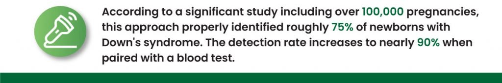
According to a significant study including over 100,000 pregnancies, this approach properly identified roughly 75% of newborns with Down’s syndrome. The detection rate increases to nearly 90% when paired with a blood test. However, the NT must be measured precisely to obtain these detection rates.
How is NT Scan performed?
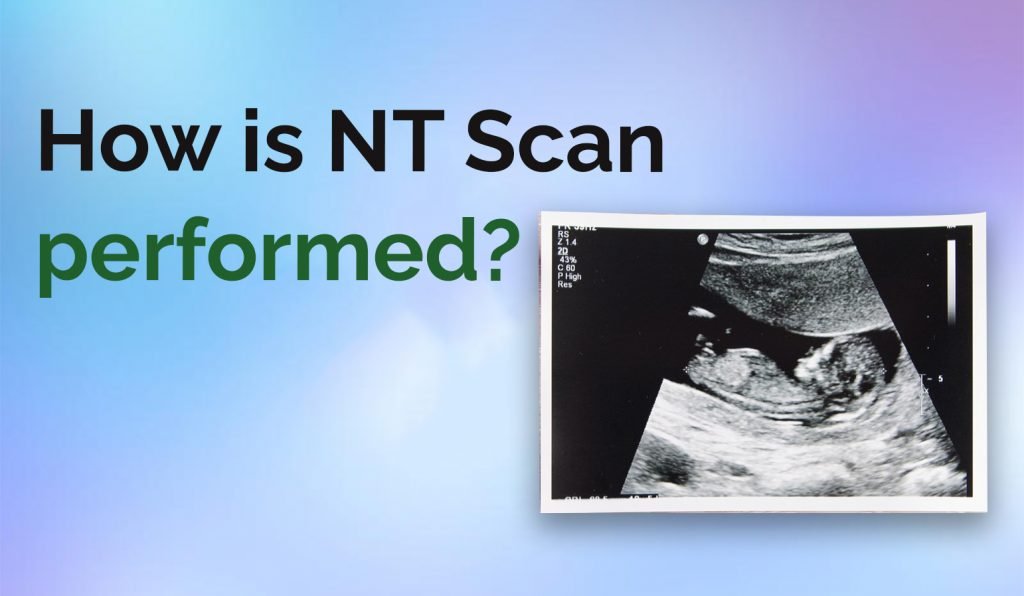
An ultrasound technician with specialized training will often perform a nuchal translucency test. However, a radiologist or an obstetrician who has obtained appropriate training to do this test may perform it.
Early pregnancy ultrasounds like the nuchal translucency scan can be performed through your abdomen or vagina. Your healthcare professional will select a method based on various factors, including the stage of your pregnancy and the shape of your body.
If your scan is done through your abdomen, you will be requested to drink a few cups of water before arriving so that your bladder is full. This facilitates viewing within your uterus (womb). Your abdomen will be softly scanned by the ultrasonic probe as some gel is applied by the sonographer. Usually, this process doesn’t hurt. If your bladder must be completely full for the test, you can let your doctor or the ultrasound technician know if it causes you any discomfort.
A tiny, lubricated ultrasound probe is gently placed into your vagina if your scan is done transvaginally. The probe is often not unpleasant, though it could be a little uncomfortable. Because the probe is closer to your uterus when scanning this technique, the images produced can be more detailed.
The results of your nuchal translucency scan can be paired with a blood test, which is often performed in weeks 10 to 12 of pregnancy, to generate a ‘combined first-trimester screen,’ or CFTS. The results of a blood test and ultrasound can provide a more accurate assessment of your risk.
